Students get through the TN Board 11th Bio Zoology Important Questions Chapter 7 Body Fluids and Circulation which is useful for their exam preparation.
TN State Board 11th Bio Zoology Important Questions Chapter 7 Body Fluids and Circulation
Answer the following.
Question 1.
In what way the circulatory system helps our body?
Answer:
The circulatory system helps to maintain the homeostasis of the body fluids and body temperature (heat exchange). The homeostatic regulation of the cardiovascular system maintains blood flow, or perfusion, to the heart and brain.
Question 2.
Why vasovagal syncope (fainting) occurs?
Answer:
Signals from the nervous system cause a sudden decrease in blood pressure, and the individual faints from lack of oxygen to the brain.
Question 3.
Name the three types of extracellular fluids.
Answer:
The three types of extracellular fluids are the interstitial fluid or tissue fluid (surrounds the cell), the plasma (fluid component of the blood) and lymph.
![]()
Question 4.
What are the four main types of plasma proteins?
Answer:
The four main types of plasma proteins synthesized in the liver are albumin, globulin, prothrombin and fibrinogen.
Question 5.
Write the functions of the plasma protein.
Answer:
Albumin maintains the osmotic pressure of the blood. Globulin facilitates the transport of ions, hormones, lipids and assists in immune function. Both Prothrombin and Fibrinogen are involved in blood clotting.
Question 6.
What happens to the constituents of blood immediately after a meal?
Answer:
The composition of plasma is not always constant. Immediately after a meal, the blood in the hepatic portal vein has a very high concentration of glucose as it is transporting glucose from the intestine to the liver where it is stored. The concentration of the glucose in the blood gradually falls after some time as most of the glucose is absorbed.
Question 7.
If too much protein consumed more than the normal need, what will happen to the excess of protein consumed?
Answer:
If too much protein is consumed, the body cannot store the excess amino acids formed from the digestion of proteins. The liver breaks down the excess amino acids and produces urea. Blood in the hepatic vein has a high concentration of urea than the blood in other vessels namely, the hepatic portal vein and hepatic artery.
![]()
Question 8.
What is the name of the pigment present in RBC? Write its important role.
Answer:
The red colour of the RBC is due to the presence of a respiratory pigment, haemoglobin dissolved in the cytoplasm. Haemoglobin plays an important role in the transport of respiratory gases and facilitates the exchange of gases with the fluid outside the cell.
Question 9.
What is haematocrit?
Answer:
Erythropoietin is a hormone secreted by the kidneys in response to low oxygen and helps in the differentiation of stem cells of the bone marrow to erythrocytes (erythropoiesis) in adults. The ratio of red blood cells to blood plasma is expressed as Haematocrit.
Question 10.
Write about the red cells of the blood.
Answer:
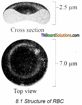
Red blood cells are abundant than other blood cells. There are about 5 million to 5.5 millions of RBC mm”3 of blood in a healthy man and 4.5 – 5.0 million RBC mnr3 in healthy women. The RBCs are very small with a diameter of about 7pm (micrometer). The structure of RBC is shown in Figure. The red colour of the RBC is due to the presence of a respiratory pigment, haemoglobin dissolved in the cytoplasm. Haemoglobin plays an important role in the transport of respiratory gases and facilitates the exchange of gases with the fluid outside the cell (tissue fluid). The biconcave shaped RBCs increases the surface area to volume ratio, hence oxygen diffuses quickly in and out of the cell. The RBCs are devoid of the nucleus, mitochondria, ribosomes and endoplasmic reticulum. The absence of these organelles accommodates more haemoglobin thereby maximising the oxygen-carrying capacity of the cell. The average life span of RBCs in a healthy individual is about 120 days after which they are destroyed in the spleen (graveyard/cemetery of RBCs) and the iron component returns to the bone marrow for reuse. Erythropoietin is a hormone secreted by the kidneys in response to low oxygen and helps in the differentiation of stem cells of the bone marrow to erythrocytes (erythropoiesis) in adults. The ratio of red blood cells to blood plasma is expressed as Haematocrit
![]()
Question 11.
Give an account of different types of white blood cell with suitable diagrams.
Answer:
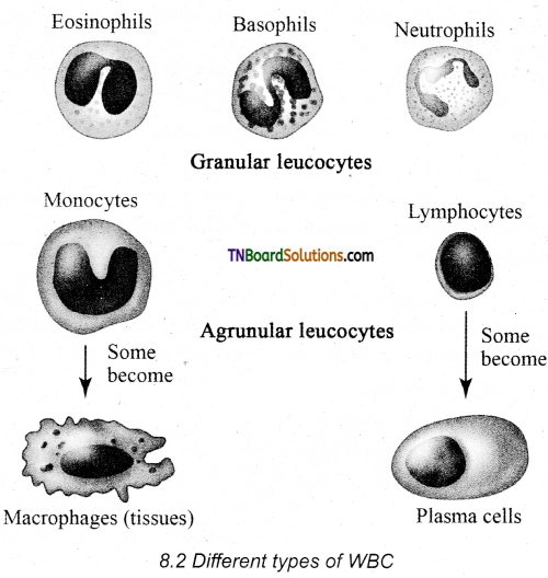
White blood cells (leucocytes) are colourless, amoeboid, nucleated cells devoid of haemoglobin and other pigments. Approximately 6000 to 8000 per cubic mm of WBCs are seen in the blood of an average healthy individual. The different types of WBCs are shown in Figure. Depending on the presence or absence of granules, WBCs are divided into two types, granulocytes and agranulocytes. Granulocytes are characterised by the presence of granules in the cytoplasm and are differentiated in the bone marrow. The granulocytes include neutrophils, eosinophils and basophils.
Neutrophils are also called heterophils or polymorphonuclear (cells with 3-4 lobes of nucleus connected with delicate threads) cells which constitute about 60% – 65% of the total WBCs. They are phagocytic in nature and appear in large numbers in and around the infected tissues.
Eosinophils have a distinctly bilobed nucleus and the lobes are joined by thin strands. They are non – phagocytic and constitute about 2 – 3% of the total WBCs. Eosinophils increase during certain types of parasitic infections and allergic reactions.
Basophils are less numerous than any other type of WBCs constituting 0.5% – 1.0% of the total number of leucocytes. The cytoplasmic granules are large-sized but fewer than eosinophils. The nucleus is large-sized and constricted into several lobes but not joined by delicate threads. Basophils secrete substances such as heparin, serotonin and histamines. They are also involved in inflammatory reactions.
Agranulocytes are characterised by the absence of granules in the cytoplasm and are differentiated in the lymph glands and spleen. These are of two types, lymphocytes and monocytes. Lymphocytes constitute 28% of WBCs. These have a large round nucleus and a small amount of cytoplasm. The two types of lymphocytes are B and T cells. Both B and T cells are responsible for the immune responses of the body. B cells produce antibodies to neutralize the harmful effects of foreign substances and T cells are involved in cell-mediated immunity.
Monocytes (Macrophages) are phagocytic cells that are similar to mast cells and have kidney shaped nucleus. They constitute 1 – 3% of the total WBCs. The macrophages of the central nervous system are the ‘microglia’, in the sinusoids of the liver they are called ‘Kupffer cells’ and in the pulmonary region they are the ‘alveolar macrophages.
Question 12.
Which type of WBC increase in its amount during allergic reactions? Write a few words about it.
Answer:
- Eosinophils increase during certain types of parasitic infections and allergic reactions.
- Eosinophils have a distinctly bilobed nucleus and the lobes are joined by thin strands. They are non – phagocytic and constitute about 2 – 3% of the total WBCs.
![]()
Question 13.
Write a few lines about the two types of lymphocytes.
Answer:
The two types of lymphocytes are B and T cells. Both B and T cells are responsible for the immune responses of the body. B cells produce antibodies to neutralize the harmful effects of foreign substances and T cells are involved in cell-mediated immunity.
Question 14.
Write short note on platelets.
Answer:
Platelets are also called thrombocytes that are produced from megakaryocytes (special cells in bone marrow) and lack nuclei. Blood normally contains 1, 50,000 – 3, 50,000 platelets mm-3 of blood. They secrete substances involved in the coagulation or clotting of blood. The reduction in platelet number can lead to clotting disorders that result in excessive loss of blood from the body.
Question 15.
Tabulate the different types of blood group in human with the antigen and antibodies.
Answer:
Distribution of antigens and antibodies in different blood groups
| Blood group | Agglutinogens (antigens) on the RBC | Agglutinin (antibodies) in the plasma |
| A | A | Anti B |
| B | B | Anti A |
| AB | AB | No antibodies |
| O | No antigens | Anti A and Anti B |
Question 16.
How can we identify the ‘+ve’ or’-ve’ factor in the blood groups?
Answer:
Rh factor is a protein (D antigen) present on the surface of the red blood cells in the majority (80%) of humans. This protein is similar to the protein present in Rhesus monkey, hence the term Rh, Individuals who carry the antigen D on the surface of the red blood cells are Rh” (Rh-positive) and the individuals who do not carry antigen D, are Rh (Rh-negative).
![]()
Question 17.
What is erythroblastosis foetalis? How is it caused? Can it be treated?
Answer:
Rh factor compatibility is also checked before blood transfusion. When a pregnant woman is Rh– and the foetus is Rh+ incompatibility (mismatch) is observed. During the first pregnancy, the Rh– antigens of the foetus does not get exposed to the mother’s blood as both their blood are separated by the placenta. However, a small amount of the foetal antigen becomes exposed to the mother’s blood during the birth of the first child. The mother’s blood starts to synthesize D antibodies. But during subsequent pregnancies, the Rh antibodies from the mother (Rh–) enters the foetal circulation and destroys the foetal RBCs. This becomes fatal to the foetus because the child suffers from anaemia and jaundice. This condition is called erythroblastosis foetalis. This condition can be avoided by the administration of anti D antibodies (Rhocum) to the mother immediately after the first childbirth.
Question 18.
Give a brief account of the general structure of blood vessels.
Answer:
The vessels carrying the blood are of three types; they are arteries, veins and capillaries. These vessels are hollow structures and have complex walls surrounding the lumen. The blood vessels in humans are composed of three layers, tunica intima, tunica media and tunica externa. The inner layer, tunica intima or tunica interna supports the vascular endothelium, the middle layer, tunica media is composed of smooth muscles and an extracellular matrix that contains a protein, elastin. The contraction and relaxation of the smooth muscles result in vasoconstriction and vasodilation. The outer layer, tunica externa or tunica adventitia is composed of collagen fibres.
Question 19.
Describe the process of clotting blood.
Answer:
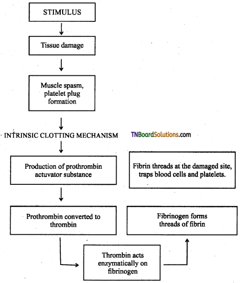
The clotting process begins when the endothelium of the blood vessel is damaged and -the connective tissue in its wall is exposed to the blood. Platelets adhere to collagen fibres in the connective tissue and release substances that form the platelet plug which provides emergency protection against blood loss. Clotting factors released from the clumped platelets or damaged cells mix with clotting factors in the plasma. The protein called prothrombin is converted to its active form called thrombin in the presence of calcium and vitamin K. Thrombin helps in the conversion of fibrinogen to fibrin threads. The threads of fibrins become interlinked into a patch that traps blood cell and seals the injured vessel until the wound is healed. After some time fibrin fibrils contract, squeezing out a straw-coloured fluid through a meshwork called serum (Plasma without fibrinogen is called serum).
![]()
Question 20.
How blood in the blood vessels are liquid in nature without clotting?
Answer:
Heparin is an anticoagulant produced in small quantities by mast cells of connective tissue which prevents coagulation in small blood vessels.
Question 21.
What are anastomoses?
Answer:
These are connections of one blood vessel (arteries) with another blood vessel. They provide an alternate route of blood flow if the original blood vessel is blocked. For example, Arteries in the joints contain numerous anastomoses. This allows blood to flow freely even if one of the arteries closes during bending of the joints.
Question 22.
What are coronary blood vessels? Why did they call so?
Answer:
Blood vessels that supply blood to the cardiac muscles with all nutrients and remove wastes are the coronary arteries and veins. Heart muscle is supplied by two arteries namely the right and left coronary arteries. These arteries are the first branch of the aorta. Arteries usually surround, the heart in the manner of a crown, hence called the coronary artery.
Question 23.
What is the Law of Laplace? Where the law can be used in the biological system?
Answer:
The Law of Laplace is used to understand the structure and function of blood vessels and the heart. Laplace law states that the tension in the walls of the blood vessel is proportional to the blood pressure and vessel radius. Blood vessels such as the aorta that is subjected to high pressures have thicker walls than the arterioles that are subjected to low pressures.
Question 24.
Why vigorous exercise after a meal can cause indigestion or abdominal cramps?
Answer:
When exercising vigorously, blood is rerouted from the digestive organ (food or no food) to the capillary beds of the skeletal muscles where it is needed immediately. This rerouting explains why vigorous exercise after a meal can cause indigestion or abdominal cramps.
![]()
Question 25.
What is incomplete double circulation?
Answer:
Amphibians have two auricles and one ventricle and no interventricular septum whereas reptiles except crocodiles have two auricles and one ventricle and an incomplete interventricular septum. Thus mixing of oxygenated and deoxygenated blood takes place in the ventricles. This type of circulation is called incomplete double circulation.
Question 26.
Write an account on the L.S. of heart with a simple sketch.
Answer:
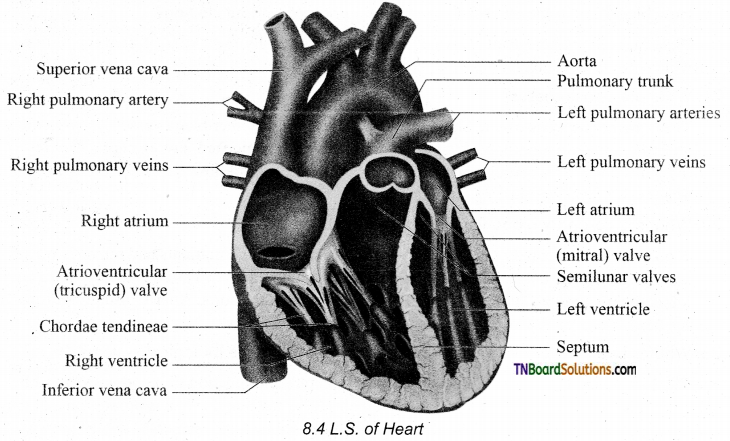
The heart is divided into four chambers, the upper two small auricles or atrium and the lower two large ventricles. The two auricles are separated by inter auricular septum and the two ventricles are separated by the interventricular septum The separation of chambers avoids mixing of oxygenated and deoxygenated blood. The auricle communicates with the ventricle through an opening called auriculo ventricular aperture which is guarded by the auriculo ventricular valves. The opening between the right atrium and the right ventricle is guarded by the tricuspid valve (three flaps or cusps), whereas* a bicuspid (two flaps or cusps) or mitral valve guards the opening between the left atrium and left ventricle. The valves of the heart allow the blood to flow only in one direction, i.e., from the atria to the ventricles and from the ventricles to the pulmonary artery or the aorta. These valves prevent the backward flow of blood. The opening of the right and left ventricles into the pulmonary artery and aorta are guarded by aortic and pulmonary valves and are called semilunar valves. The opening and closing of the semilunar valves are achieved by the chordae tendinae. The chordae tendinae are attached to the lower end of the heart by papillary muscles.
Question 27.
What does the following term define?
- Heartbeat,
- Systole,
- Diastole,
- ‘lub’ and ‘dub’ sound of the heart,
- Cardiac cycle.
Answer:
- Rhythmic contraction and expansion of the heart are called heartbeat.
- The contraction of the heart is called systole.
- The relaxation of the heart is called diastole.
- The first heart sound (lub) is associated with the closure of the tricuspid and bicuspid valves whereas the second heart sound (dub) is associated with the closure of the semilunar valves.
- The events that occur at the beginning of the heartbeat and last until the beginning of the next beat is called the cardiac cycle.
![]()
Question 28.
Write about the series of event takes place in the cardiac cycle.
Answer:
The series of events takes place in a cardiac cycle.
PHASE 1: Ventricular diastole – The pressure in the auricles increases than that of the ventricular pressure. AV valves are open while the semilunar valves are closed. Blood flows from the auricles into the ventricles passively.
PHASE 2: Atrial systole – The atria contracts while the ventricles are still relaxed. The contraction of the auricles pushes the maximum volume of blood to the ventricles until they reach the end-diastolic volume (EDV). EDV is related to the length of the cardiac muscle fibre. The more the muscle is stretched, greater the EDV and the stroke volume.
PHASE 3: Ventricular systole (isovolumetric contraction) – The ventricular contraction forces the AV valves to close and increases the pressure inside the ventricles. The blood is then pumped from the ventricles into the aorta without a change in the size of the muscle fibre length and ventricular chamber volume (isovolumetric contraction).
PHASE 4: Ventricular systole (ventricular ejection) – Increased ventricular pressure forces the semilunar valves to open and blood is ejected out of the ventricles without backflow of blood. This point is the end of the systolic volume (ESV).
PHASE 5: (Ventricular diastole) – The ventricles begin to relax, pressure in the arteries exceeds ventricular pressure, resulting in the closure of the semilunar valves. The heart returns to phase 1 of the cardiac cycle.
Question 29.
Define cardiac output. How can it be calculated?
Answer:
The amount of blood pumped out by each ventricle per minute is called cardiac output(CO).
It is a product of heart rate (HR) and stroke volume (SV). Heart rate or pulse is the number of beats per minute.
Pulse pressure = systolic pressure – diastolic pressure.
Stroke volume (SV) is the volume of blood pumped out by one ventricle with each beat. SV depends on ventricular contraction.
CO = HR x SV
SV represents the difference between EDV (amount of blood that collects in a ventricle during diastole) and ESV (volume of blood remaining in the ventricle after contraction).
SV = EDV – ESV
Question 30.
Explain how the Frank-Starling effect protects the heart from an abnormal increase in blood volume.
Answer:
According to the Frank-Starling law of the heart, the critical factor controlling SV is the degree to which the cardiac muscle cells are stretched just before they contract. The most important factor stretching the cardiac muscle is the amount of blood returning to the heart and distending its ventricles, venous return. During vigorous exercise, SV may double as a result of venous return. The heart’s pumping action normally maintains a balance between cardiac output and venous return. Because the heart is a double pump, each side can fail independently of the other. If the left side of the heart fails, it results in pulmonary congestion and if the right side fails, it results in peripheral congestion. Frank-Starling effect protects the heart from an abnormal increase in blood volume.
![]()
Question 31.
What is blood pressure? What are its types? How is it caused?
Answer:
- Blood pressure is the pressure exerted on the surface of blood vessels by the blood.
- There are two types of pressure, systolic pressure and diastolic pressure.
- Systolic pressure is the pressure in the arteries as the chambers of the heart contracts. Diastolic pressure is the pressure in the arteries when the heart chambers relax.
Question 32.
What is orthostatic hypotension?
Answer:
The primary reflex pathway for homeostatic control of mean arterial pressure is the baroreceptor reflex. The baroreceptor reflex functions every morning when you get out of bed. When you are lying flat the gravitational force is evenly distributed. When you stand up, gravity causes blood to pool in the lower extremities. The decrease in blood pressure upon standing is known as orthostatic hypotension.
Question 33.
What is ECG? How ECG graph helps the activity of the heart during one cardiac cycle?
Answer:
Electrocardiogram (ECG): An electrocardiogram (ECG) records the electrical activity of the heart over a period of time using electrodes placed on the skin, arms, legs and chest.
It records the changes in electrical potential across the heart during one cardiac cycle.
A normal ECG shows 3 waves designated as P wave, QRS complex and T wave.
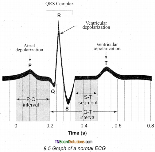
P Wave (atrial depolarisation): It is a small upward wave and indicates the depolarization of the atria. This is the time taken for the excitation to spread through the atria from the SA node. Contraction of both atria lasts for around 0.8 – 1.0 sec.
PQ Interval (AV node delay): It is the onset of the P wave to the onset of the QRS complex. This is from the start of depolarization of the atria to the beginning of ventricular depolarization. It is the time taken for the impulse to travel from the atria to the ventricles (0.12 – 0.21 sec). It is the measure of AV conduction time.
QRS Complex (ventricular depolarisation): No separate wave for atrial depolarization in the ECG is visible. Atrial depolarization occurs simultaneously with ventricular depolarization. The normal QRS complex lasts for 0.06 – 0.09 sec. The QRS complex is shorter than the P wave because depolarization spreads through the Purkinje fibres. Prolonged QRS wave indicates delayed conduction through the ventricle, often caused due to ventricular hypertrophy or due to a block in the branches of the bundle of His.
ST-Segment: It lies between the QRS complex and the T wave. It is the time during which all regions of the ventricles are completely depolarised and reflects the long plateau phase before repolarisation. In the heart muscle, prolonged depolarization is due to retardation of K+ efflux and is responsible for the plateau. The ST segment lasts for 0.09 sec.
T wave (ventricular depolarisation): It represents ventricular depolarization. The duration of the T wave is longer than the QRS complex because repolarisation takes place simultaneously throughout the ventricular depolarisation.
![]()
Question 34.
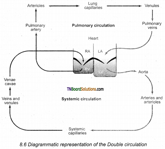
Show the double circulation by way of the diagrammatic sketch.
Answer:
Question 35.
In what way systemic circulation is different from pulmonary circulation incomplete double blood circulation?
Answer:
In the systemic circulation, the oxygenated blood entering the aorta from the left ventricle is carried by a network of arteries, arterioles and capillaries to the tissues. The deoxygenated blood from the tissue is collected by venules, veins and vena cava and emptied into the right atrium. In pulmonary circulation, the blood from the heart (right ventricle) is taken to the lungs by the pulmonary artery and the oxygenated blood from the lungs is emptied into the left auricle by the pulmonary vein.
Question 36.
How cardiac activity is regulated?
Answer:
The type of heart in human is myogenic because the heartbeat originates from the muscles of the heart. The nervous and endocrine systems work together with paracrine signals (metabolic activity) to influence the diameter of the arterioles and alter the blood flow. Neuronal control is achieved through the autonomic nervous system (sympathetic and parasympathetic). Sympathetic neurons release norepinephrine and the adrenal medulla releases epinephrine. The two hormones bind to p – adrenergic receptors and increase the heart rate. The parasympathetic neurons secrete acetylcholine that binds to muscarinic receptors and decreases the heartbeat.
Question 37.
Write about the following disorders of the circulatory system.
(a) Hypertension
(b) Coronary heart disease.
(c) Angina pectoris.
(d) Stroke
(e) Myocardial infarction.
(f) Rheumatoid heart disease.
Answer:
Hypertension is the most common circulatory disease. The normal blood pressure in man is 120/80 mm Hg. In cases when the diastolic pressure exceeds 90 mm Hg and the systolic pressure exceeds 150 mm Hg persistently, the condition is called hypertension. Uncontrolled hypertension may damage the heart, brain and kidneys.
Coronary heart disease occurs when the arteries are lined by atheroma. The build-up of atheroma contains cholesterol, fibres, dead muscle and platelets and is termed Atherosclerosis. The cholesterol-rich atheroma forms plaques in the inner lining of the arteries making them less elastic and reduces the blood flow. Plaque grows within the artery and tends to form blood clots, forming coronary thrombus. A thrombus in a coronary artery results in a heart attack.
Angina pectoris (ischemic pain in the heart muscles) is experienced during the early stages of coronary heart disease. Atheroma may partially block the coronary artery and reduce the blood supply to the heart. As a result, there is tightness or choking with difficulty in breathing. This leads to angina or chest pain. Usually, it lasts for a short duration of time.
Stroke is a condition when the blood vessels in the brain bursts, (Brain haemorrhage) or when there is a block in the artery that supplies the brain, (atherosclerosis) or thrombus. The part of the brain tissue that is supplied by this damaged artery dies due to a lack of oxygen (cerebral infarction).
Myocardial infarction (Heart failure): The prime defect in heart failure is a decrease in cardiac muscle contractility. The Frank-Starling curve shifts downwards and towards the right such that for a given EDV, a failing heart pumps out a smaller stroke volume than a normal healthy heart.
When the blood supply to the heart muscle or myocardium is remarkably reduced it leads to the death of the muscle fibres. This condition is called a heart attack or myocardial infarction. The blood clot or thrombosis blocks the blood supply to the heart and weakens the muscle fibres. It is also called Ischemic heart disease due to a lack of oxygen supply to the heart muscles. If this persists it leads to chest pain or angina. Prolonged angina leads to death of the heart muscle resulting in heart failure.
Rheumatoid Heart Disease: Rheumatic fever is an autoimmune disease that occurs 2-4 weeks after throat infection usually a streptococcal infection. The antibodies developed to combat the infection cause damage to the heart. Effects include fibrous nodules on the mitral valve, fibrosis of the connective tissue and accumulation of fluid in the pericardial cavity.
![]()
Question 38.
What is CPR? Which case is more useful?
Answer:
CPR is a life-saving procedure that is done at the time of emergency conditions such as when a person’s breath or heartbeat has stopped abruptly in case of drowning, electric shock or heart attack. CPR includes rescue of breath, which is achieved by mouth to mouth breathing, to deliver oxygen to the victim’s lungs by external chest compressions which helps to circulate blood to the vital organs. CPR must be performed within 4 to 6 minutes after cessation of breath to prevent brain damage or death.
Question 39.
How varicose veins are formed? Where are they usual located?
Answer:
Varicose veins The veins are so dilated that the valves prevent the backflow of blood. The veins lose their elasticity and become congested. Common sites are legs, rectoanal regions (haemorrhoids), the oesophagus and the spermatic cord.
Question 40.
What is embolism?
Answer:
Embolism is the obstruction of the blood vessel by an abnormal mass of materials such as a fragment of the blood clot, bone fragment or an air bubble. An embolus may lodge in the lungs, coronary artery or liver and leads to death.
Question 41.
What is an aneurysm? How much it is dangerous?
Answer:
Aneurysm the weakened regions of the wall of the artery or veins bulges to form a balloon-like sac. An unruptured aneurysm may exert pressure on the adjacent tissues or may burst to cause a massive haemorrhage.
Question 42.
Mention the three layers of blood vessels.
Answer:
The blood vessels in humans are composed of three layers, tunica intima, tunica media and tunica externa.
Choose the correct answer.
1. The average blood volume in an adult is:
(a) 5000 ml
(b) 50000 ml
(c) 50 ml
(d) 5000 l
Answer:
(a) 5000 ml
![]()
2. Liver receives oxygenated blood from the heart through:
(a) hepatic portal vein
(b) hepatic vein
(c) hepatic artery
(d) Renal artery
Answer:
(c) hepatic artery
3. ………… plasma protein is responsible for maintaining the osmotic pressure of blood.
(a) Globulin
(b) Prothrombin
(c) Fibrinogen
(d) Albumin
Answer:
(d) Albumin
4. ………… plasma proteins are involved in blood clotting.
(a) Albumin and prothrombin
(b) Globulin and albumin
(c) Prothrombin and fibrinogen
(d) Globulin and fibrinogen.
Answer:
(c) Prothrombin and fibrinogen
5. Healthy man has ………. millions of RBC in mm-3 of blood.
(a) 5.0-5.5
(b) 5.0-6
(c) 4.5-5.0
(d) 5.5-5.6
Answer:
(a) 5.0-5.5
6. Healthy women has ……….. millions of RBC in mm-3 of blood.
(a) 5.0 – 5.5
(b) 5.5 – 6.0
(c) 4.5 – 5.5
(d) 4.5 – 5.0
Answer:
(d) 4.5 – 5.0
7. Human RBC has ………… number of nucleus.
(a) 3
(b) 2
(c) 0
(d) 1
Answer:
(c) 0
8. The average life span of RBCs in a healthy individual is:
(a) 102 days
(b) 201 days
(c) 120 days
(d) 21 days.
Answer:
(c) 120 days
![]()
9. After the life span of RBCs are over they get destroyed in the:
(a) kidneys
(b) bone marrow
(c) thymus
(d) spleen
Answer:
(d) spleen
10. Differentiation of stem cells of the bone marrow to erythrocytes in adults is called:
(a) erythropoiesis
(b) apoptosis
(c) coagulation
(d) embolus
Answer:
(a) erythropoiesis
11. ………….. are the types of WBCs which are involved in inflammatory reactions.
(a) Neutrophils
(b) Basophils
(c) Eosinophils
(d) Monocytes
Answer:
(b) Basophils
12. The macrophages of the liver are called:
(a) microglia
(b) alveolar macrophages
(c) kupffer cells
(d) lymphocytes
Answer:
(c) kupffer cells
13. Blood normally contains ……….. platelets mm-3 of blood.
(a) 1,00,000 – 2,00,000
(b) 1,50,000 – 3,50,000
(c) 3,50,000 – 4,00,000
(d) 2,50,000 – 3,50,000
Answer:
(b) 1,50,000 – 3,50,000
14. The Rh factor protein is otherwise called ………… present on the surface of the red blood cells, in the majority of human.
(a) B – antigen
(b) D – antigen
(c) C – antigen
(d) A – antigen
Answer:
(b) D – antigen
![]()
15. The capillaries doesn’t posses the ………… layer of the blood vessel.
(a) tunica media
(b) tunica intima
(c) tunica Externa
(d) epicardium
Answer:
(a) tunica media
16. Complete double circulation is seen in ………….
(a) amphibians
(b) crocodiles
(c) fishes
(d) annelids
Answer:
(b) crocodiles
17. ………… valve of the heart is otherwise called mitral valve.
(a) Semilunar valves
(b) Tricuspid valves
(c) Bicuspid valves
(d) Atrial valve
Answer:
(c) Bicuspid valves
18. The heart sounds can be heard through …………
(a) electrocardiogram
(b) sphygmomanometer
(c) haemocytometer
(d) stethoscope
Answer:
(d) stethoscope
19. The amount of blood pumped out by each ventricle per minute is called:
(a) pulse pressure
(b) cardiac output
(c) stroke volume
(d) heart rate.
Answer:
(b) cardiac output
20. The volume of blood pumped out by one ventricle with each beat is called:
(a) Cardiac output
(b) Heartbeat
(c) Stroke volume
(d) systolic pressure
Answer:
(c) Stroke volume
![]()
21. Stroke volume depends on:
(a) Ventricular contraction
(b) Ventricular relaxation
(c) Atrial contraction
(d) Atrial relaxation
Answer:
(a) Ventricular contraction
22. Blood pressure of man is measured by using:
(a) Electrocardiogram
(b) Sphygmomanometer
(c) Cytometer
(d) Computed tomography
Answer:
(b) Sphygmomanometer
23. Normal blood pressure in man is:
(a) 120/80 mm Hg
(b) 130/70 mm Hg
(c) 120/60 mm Hg
(d) 150/60 mm Hg
Answer:
(a) 120/80 mm Hg
24. The QRS complex wave of ECG lasts for ………… seconds.
(a) 0.05 -0.1
(b) 0.06 – 0.09
(c) 0.8
(d) 0.12-0.21
Answer:
(b) 0.06 – 0.09
25. The blood clot inside the blood vessel is called:
(a) thrombus
(b) embolus
(c) atheroma
(d) plaques
Answer:
(b) embolus
![]()
26. Cells found in the lymphatics are called:
(a) basophils
(b) neutrophils
(c) lymphocytes
(d) monocytes
Answer:
(c) lymphocytes
27. Plasma without fibrinogen is called:
(a) Plasma protein
(b) Serum
(c) Fibrin
(d) atheroma
Answer:
(b) Serum
28. The damaged tissues and dead leucocytes are removed from the body in the forms of:
(a) pus
(b) blood
(c) serum
(d) plasma
Answer:
(a) pus
29. Match:
| Column – I | Column II |
| (i) Kupffer cells | (a) Heterophils |
| (ii) Neutrophils | (b) Ratio of RBC to blood plasma |
| (iii) Platelets | (c) Liver |
| (iv) White blood cells | (d) Thrombocytes |
| (v) Haematocrit | (e) Leucocytes |
(a) (i)-(c); (ii)-(a); (iii)-(d); (iv)-(e); (v)-(b)
(b) (i)-(d); (ii)-(a); (iii)-(b); (iv)-(e); (v)-(c)
(c) (i)-(c); (ii)-(b); (iii)-(d); (iv)-(a); (v)-(e)
(d) (i)-(a); (ii)-(c); (iii)-(e); (iv)-(b); (v)-(d)
Answer:
(a) (i)-(c); (ii)-(a); (iii)-(d); (iv)-(e); (v)-(b)