Students get through the TN Board 11th Bio Zoology Important Questions Chapter 10 Neural Control and Coordination which is useful for their exam preparation.
TN State Board 11th Bio Zoology Important Questions Chapter 10 Neural Control and Coordination
Answer the following.
Question 1.
What are the basic functions of the neural systems in higher animals?
Answer:
The neural system of higher animals is well developed and performs the following basic functions:
- Sensory functions: It receives sensory input from the internal and external environments.
- Motor functions: It transmits motor commands from the brain to the skeletal and muscular systems.
- Autonomic functions: Reflex actions.
Question 2.
What are the divisions of the human neural system?
Answer:
The human neural system is divided into two, the central neural system (CNS) and the peripheral neural system.
Question 3.
What are the divisions of neurons based on their function?
Answer:
There are three functional classes of neurons. They are the afferent neurons that take sensory impulses to the Central Neural system (CNS) from the sensory organs; the efferent neurons that carry motor impulses from the CNS to the effector organs; and interneurons that lie entirely within the CNS between the afferent and efferent neurons.
![]()
Question 4.
Write the functions performed by Neuroglia.
Answer:
Neuroglia performs several functions such as providing nourishment to the surrounding neurons; involving, the memory process; repairing the injured tissues due to their dividing and regenerating capacity; and acting as phagocyte cells to engulf the foreign particles at the time of any injury to the brain.
Question 5.
Describe the structure of a neuron, which is the functional unit of the nervous system with a neat sketch.
Answer:
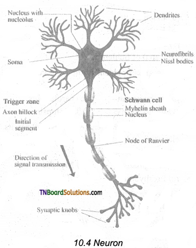
A neuron is a microscopic structure composed of three major parts namely the cell body (soma), dendrites, and axon. The cell body is the spherical part of the neuron that contains all the cellular organelles as a typical cell (except centriole). The plasma membrane covering the neuron is called neurilemma and the axon is axolemma. The repeatedly branched short fibers coming out of the cell body are called dendrites, which transmit impulses towards the cell body. The cell body and the dendrites contain cytoplasm and granulated endoplasmic reticulum called Nissl’s granules.
An axon is a long fiber, that arises from a cone-shaped area of the cell body called the Axon hillock and ends at the branched distal end.
Axon hillock is the place where the nerve impulse is generated in the motor neurons.
The axon of one neuron branches and forms connections with many other neurons. An axon contains the same organelles found in the dendrites and cell body but lacks Nissl’s granules and Golgi apparatus.
The axon, particularly of peripheral nerves is surrounded by Schwann cells to form the myelin sheath, which acts as an insulator.
Myelin sheath is associated only with the axon; dendrites are always non-myelinated. Schwann cells are not continuous along the axon; so there are gaps in the myelin sheath between adjacent Schwann cells. These gaps are called Nodes of Ranvier. Large myelinated nerve fibers conduct impulses rapidly, whereas non-myelinated fibers conduct impulses quite slowly.
Each branch at the distal end of the axon terminates into a bulb-like structure called a synaptic knob which possesses synaptic vesicles filled with neurotransmitters. The axon transmits nerve impulses away from the cell body to the interneural space or to a neuromuscular junction.
![]()
Question 6.
Classify neurons according to their structural difference with a simple diagram.
Answer:
The neurons are divided into three types based on the number of axons and dendrites they possess.
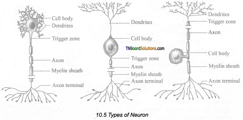
- Multipolar neurons have many processes with one axon and two or more dendrites. They are mostly interneurons.
- Bipolar neurons have two, processes with one axon and one dendrite. These are found in the retina of the eye, inner ear, and the olfactory area of the brain.
- Unipolar neurons have a single short process and one axon. Unipolar neurons are located in the ganglia of cranial and spinal nerves.
Question 7.
What are the two phases of transmission of nerve impulses?
Answer:
The transmission of impulse involves two main phases:
- Resting membrane potential,
- Action membrane potential.
Question 8.
Write about the “resting membrane potential” state of impulse transmission.
Answer:
Resting membrane Potential: The electrical potential difference across the plasma membrane of a resting neuron is called the resting potential during which the interior of the cell is negative due to greater efflux of K+ outside the cell than Na+ influx into the cell. When the axon is not conducting any impulses i.e. in resting condition, the axon membrane is more permeable to K+ and less permeable to Na+ ions, whereas it remains impermeable to negatively charge protein ions.
The axoplasm contains a high concentration of K+ and negatively charged proteins and a low concentration of Na+ ions. In contrast, fluid outside the axon (ECF) contains a low concentration of K+ and a high concentration of Na+, and this forms a concentration gradient. This ionic gradient across the resting membrane is maintained by ATP driven Sodium-Potassium pump, which exchanges 3Na+ outwards for 2K+ into the cells. In this state, the cell membrane is said to be polarized. In neurons, the resting membrane potential ranges from – 40mV to – 90mV, and its normal value is – 70mV. The minus sign indicates that the inside of the cell is negative with respect to the outside.
![]()
Question 9.
Write briefly about the different phases of action membrane potential.
Answer:
Action membrane potential: An action potential occurs when a neuron sends information down an axon, away from the cell body. It includes the following phases, depolarization, repolarization, and hyperpolarization.
Depolarization – Reversal of polarity: When a nerve fiber is stimulated, sodium voltage-gated opens and makes the axolemma permeable to Na+ ions; meanwhile the potassium voltage gate closes. As a result, the rate of flow of Na+ ions into the axoplasm exceeds the rate of flow of K+ ions to the outside fluid [ECF]. Therefore, the axolemma becomes positively charged inside and negatively charged outside. This reversal of electrical charge is called Depolarization.
During depolarization, when enough Na+ ions enter the cell, the action potential reaches a certain level, called threshold potential [-55mV], The particular stimulus. which is able to bring the membrane potential to the threshold is called threshold stimulus.
The action potential occurs in response to a threshold stimulus but does not occur at subthreshold stimuli. This is called the all or none principle. Due to the rapid influx of Na+ ions, the membrane potential shoots rapidly up to +45mV which is called the Spike potential.
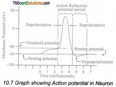
Repolarisation [Falling Phase]: When the membrane reaches the spike potential, the sodium voltage-gated closes and potassium voltage-gated opens. It checks the influx of Na+ ions and initiates the efflux of K+ ions which lowers the number of positive ions within the cell. Thus, the potential falls back towards the resting potential. The reversal of membrane potential inside the axolemma to negative occurs due to the efflux of K+ ions. This is called Repolarisation.
Hyperpolarization: If repolarization becomes more negative than the resting potential -70 mV to about -90 mV, it is called Hyperpolarization. During this, K+ ion gates are more permeable to K+ even after reaching the threshold level as it closes slowly; hence called Lazy gates. The membrane potential returns to its original resting state when K+ ion channels close completely. During hyperpolarization, the Na+ voltage gate remains closed.
Question 10.
Is there any speed difference among the neurons? Why there is such a difference?
Answer:
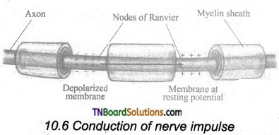
The conduction speed of a nerve impulse depends on the diameter of the axon. The greater the axon’s diameter, the faster is the conduction. The myelinated axon conducts the impulse faster than the non-myelinated axon.
![]()
Question 11.
What is ‘saltatory conduction?
Answer:
The voltage-gated Na+ and K+ channels are concentrated at the nodes of Ranvier. As a result, the impulse jumps node to node, rather than traveling the entire length of the nerve fiber. This mechanism of conduction is called Saltatory Conduction.
Question 12.
How nerve impulse is transmitted in the synaptic region of the neurons?
Answer:
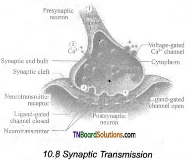
The junction between two neurons is called a Synapse through which a nerve impulse is transmitted. The first neuron involved in the synapse forms the pre-synaptic neuron and the second neuron is the post-Synaptic neuron. A small gap between the pre and postsynaptic membranes is called Synaptic Cleft that forms a structural gap and a functional bridge between neurons. The axon terminals contain synaptic vesicles filled with neurotransmitters. When an impulse [action potential] arrives at the axon terminals, it depolarizes the presynaptic membrane, opening the voltage-gated calcium channels. The influx of calcium ions stimulates the synaptic vesicles towards the pre-synaptic membrane and fuses with it. In the neurilemma, the vesicles release their neurotransmitters into the synaptic cleft by exocytosis. The released neurotransmitters bind to their specific receptors on the post¬synaptic membrane, responding to chemical signals. The entry of the ions can generate a new potential in the post-synaptic neuron, which may be either excitatory or inhibitory. Excitatory post-synaptic potential causes depolarization whereas inhibitory post-synaptic potential causes hyperpolarization of postsynaptic membrane.
Question 13.
What are the three meninges of the brain? Where are they present.
Answer:
The brain is located in the cranial cavity and is covered by three cranial meninges.
- The outer thick layer is Duramater which lines the inner surface of the cranial cavity.
- The median thin layer is the Arachnoid mater which is separated from the dura mater by a narrow subdural space.
- The innermost layer is Piamater which is closely adhered to the brain but separated from the arachnoid mater by the subarachnoid space.
![]()
Question 14.
Write an account of the structure and function of the forebrain.
Answer:
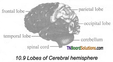
Fore Brain, comprises the following regions: Cerebrum and Diencephalon. The cerebrum is the ‘seat of intelligence’ and forms the major part of the brain. The cerebrum consists of an outer cortex, inner medulla, and basal nuclei. The superficial region of the cerebrum is called the cerebral cortex, which looks grey due to the presence of unmyelinated nerve cells. The cerebral cortex consists of the neuronal cell body, dendrites, associated glial, and blood vessels. The surface of the cerebrum shows many convolutions (folds) and grooves. The folds are called gyri (singular gyrus); the shallow grooves between the gyri are called sulci (singular sulcus) and deep grooves are called fissures. These sulci and gyri increase the surface area of the cerebral cortex. Several sulci divide the cerebrum into eight lobes: a pair of frontals, parietals, temporals, and occipital lobes.
A median longitudinal fissure divides the cerebrum longitudinally into two cerebral hemispheres. A transverse fissure separates the cerebral hemispheres from the cerebellum. The hemispheres are connected by a tract of nerve fibers called the corpus callosum. The cerebral cortex has three functional areas namely sensory areas that occur in the parietal, temporal, and occipital lobes of the cortex. They receive and interpret the sensory impulses. The motor area of the cortex which controls voluntary muscular movements lies in the posterior part of the frontal lobes. The areas other than sensory and motor areas are called Association areas that deal with integrative functions such as memory, communications, learning, and reasoning. Inner to the cortex is the medulla which is white in color and acts as a nerve tract between the cortex and the diencephalon.
Diencephalon consists largely of the following three paired structures.
The epithalamus forms the roof of the diencephalon and it is a non-nervous tissue. The anterior part of the epithalamus is vascular and folded to form the choroid plexus. Just behind the choroid plexus, the epithalamus forms a short stalk that ends in a rounded body called the pineal body which secretes the hormone, melatonin which regulates the sleep and wake cycle.
Thalamus is composed of grey mater which serves as a relay center for impulses between the spinal cord, brain stem, and cerebrum. Within the thalamus, information is sorted and edited and plays a key role in learning and memory. It is a major coordinating center for sensory and motor signaling.
Hypothalamus forms the floor of the diencephalon. The downward extension of the hypothalamus, the infundibulum connects the hypothalamus with the pituitary gland. The hypothalamus contains a pair of small rounded bodies called mammillary bodies that are involved in olfactory reflexes and emotional responses to odor. Hypothalamus maintains homeostasis and has many centers which control the body temperature, urge for eating, and drinking. It also contains a group of neurosecretory cells that secrete the hypothalamic hormones. Hypothalamus also acts as the satiety center.
Functions of brain lobes
| Structure | Functions |
| Frontal | Behaviour, Intelligence, Memory, Movement |
| Parietal | Language, Reading, Sensation |
| Temporal | Speech, Hearing, Memory |
| Occipital | Visual processing |
Question 15.
What is corpora quadrigemina? What is its function?
Answer:
The dorsal portion of the midbrain consists of four rounded bodies called corpora quadrigeminal which acts as a reflex center for vision and hearing.
![]()
Question 16.
Given an account of the different areas of the hindbrain region.
Answer:
Hindbrain: Rhombencephalon forms the hindbrain. It comprises of cerebellum, pons varolii and medulla oblongata. The cerebellum is the second largest part of the brain. It consists of two cerebellar hemispheres and a central worm-shaped part, the vermis. The cerebellum controls and coordinates muscular movements and body equilibrium. Any damage to the cerebellum often results in uncoordinated voluntary muscle movements.
Pons varoli lies infront of the cerebellum between the midbrain and the medulla oblongata. The nerve fibers in the pons Varolii form a bridge between the two cerebellar hemispheres and connect the medulla oblongata with the other region of the brain. The respiratory nuclei found in the pons cooperate with the medulla to control respiration.
Medulla oblongata forms the posterior-most part of the brain. It connects the spinal cord with various parts of the brain. It receives and integrates signals from the spinal cord and sends it to the cerebellum and thalamus. The medulla contains vital centers that control cardiovascular reflexes, respiration, and gastric secretions.
Question 17.
What are the fluid-filled spaces of the brain called? Write few lines about its position.
Answer:
Ventricles of the Brain: The brain has four hollow, fluid-filled spaces. The C-shaped space found inside each cerebral hemisphere forms the lateral ventricles I and II which are separated from each other by a thin membrane called the septum pellucidum. Each lateral ventricle communicates with the narrow III ventricle in the diencephalon through an opening called interventricular foramen (foramen of Monro). The ventricle III is continuous with the ventricle IV in the hindbrain through a canal called the aqueduct of Sylvius.
Question 18.
Brief an account on the G.S. of the spinal cord with a simple sketch.
Answer:
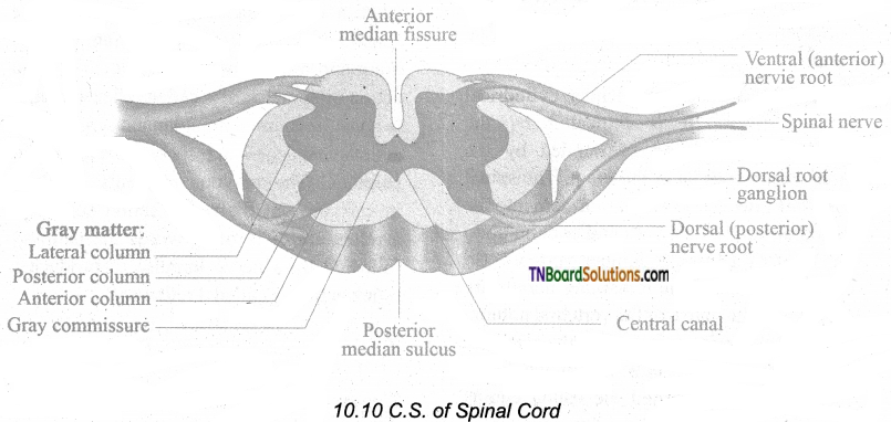
In the cross-section of the spinal cord, there are two indentations: the posterior median sulcus and the anterior median fissure. Although there might be slight variations, the cross-section of the spinal cord is generally the same
throughout its length. In contrast to the brain, the grey matter in the spinal cord forms an inner butterfly-shaped region surrounded by the outer white matter. The grey matter consists of neuronal cell bodies and their dendrites, interneurons, and glial cells. White matter consists of bundles of nerve fibers. In the center of the grey matter, there is a central canal that is filled with CSF. Each half of the grey matter is divided into a dorsal horn, a ventral horn, and a lateral horn.
The dorsal horn contains cell bodies of interneurons on which afferent neurons terminate. The ventral horn contains cell bodies of the efferent motor neurons supplying the skeletal muscle. Autonomic nerve fibers, supplying cardiac and smooth muscles and exocrine glands, originate from the cell bodies found in the lateral horn. In the white matter, the bundles of nerve fibers form two types of tracts namely ascending tracts which carry sensory impulses to the brain, and descending tracts which carry motor impulses from the brain to the spinal nerves at various levels of the spinal cord. The spinal cord shows two enlargements, one in the cervical region and another one in the lumbosacral region. The cervical enlargement serves the upper limb and lumbar enlargement serves the lower limbs.
![]()
Question 19.
Write the anatomical structure of the spinal cord.
Answer:
The spinal cord is a long, slender, cylindrical nervous tissue. It is protected by the vertebral column and surrounded by the three membranes as in the brain. The spinal cord extends from the brain stem into the vertebral canal of the vertebral column up to the level of 1st or 2nd lumbar vertebra. So the nerve roots of the remaining nerves are greatly elongated to exit the vertebral column at their appropriate space. The thick bundle of elongated nerve roots within the lower vertebral canal is called the cauda equina (horse’s tail) because of its appearance.
Question 20.
In what way the grey arid white matter of the brain and the spiral cord differs.
Answer:
The superficial region of the cerebrum is called the cerebral cortex, which looks grey due to the presence of unmyelinated nerve cells. The cerebral cortex consists of the neuronal cell body, dendrites, associated glial, and blood vessels. Inner to the cortex is the medulla which is white in color and acts as a nerve tract between the cortex and the diencephalon. In contrast to the brain, the grey matter in the spinal cord forms an inner butterfly-shaped region surrounded by the outer white matter. The grey matter consists of neuronal cell bodies and their dendrites, interneurons, and glial cells. White matter consists of bundles of nerve fibers. In the center of the grey matter, there is a central canal that is filled with CSF.
Question 21.
On touching a hot pan, the hand is withdrawn rapidly without our willingness. What is this action called? Which part is controlling this action? How it is occurs?
Answer:
On touching a hot pan, the hand is withdrawn rapidly. This is called reflex action.
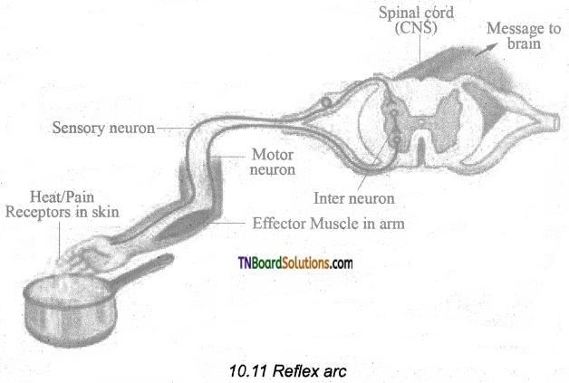
The spinal cord remains as a connecting functional nervous structure in between the brain and effector organs. But sometimes when a very quick response is needed, the spinal cord can affect motor initiation as the brain and brings about an effect. This rapid action by the spinal cord is called reflex action. It is a fast, involuntary, unplanned sequence of actions that occurs in response to a particular stimulus. The nervous elements involved in carrying out the reflex action constitute a reflex arc or in other words, the pathway followed by a nerve impulse to produce a reflex action is called a reflex arc.
Question 22.
Draw the schematic sketch of functional components of a reflex arc.
Answer:
Sensory Receptor: It is a sensory structure that responds to a specific stimulus.
Sensory Neuron: This neuron takes the sensory impulse to the grey (afferent) matter of the spinal cord through the dorsal root of the spinal cord.
Interneurons: One or two interneurons may f serve to transmit the impulses from the d sensory neuron to the motor neuron.
Motor Neuron: It transmits impulses from CNS to the effector organ.
Effector Organs: It may be a muscle or gland which responds to the impulse received.
Sensory organ → Sensory of afferent neuron → Grey matter of the spinal cord → Intermediary or relay neuron → efferent or motor neuron → effector organ.
![]()
Question 23.
Write about the types of reflexes.
Answer:
There are two types of reflexes. They are –
- Unconditional reflex is an inborn reflex for an unconditioned stimulus. It does not need any past experience, knowledge, or training to occur; eg: Blinking of an eye when a dust particle about to fall into it, sneezing, and coughing due to foreign particles entering the nose or larynx.
- A conditioned reflex is a response to a stimulus that has been acquired by learning. This does not naturally exist in animals. Only an experience makes it a part of the behavior, eg: The excitement of the salivary gland on seeing and smelling food. The conditioned reflex was first demonstrated by the Russian physiologist Pavlov in his classical conditioning experiment in a dog. The cerebral cortex controls the conditioned reflex.
Question 24.
What is the peripheral neural system? What are its components?
Answer:
- Peripheral Neural System (PNS) consists of all nervous tissue outside the CNS.
- Components of PNS include nerves, ganglia, enteric plexuses, and sensor receptors.
Question 25.
Write out cranial nerves rich are arising from the brain coming under the peripheral neural system.
Answer:
Cranial nerves: There are 12 pairs of cranial nerves, of which the first two pairs arise from the forebrain and the remaining 10 pairs from the midbrain. Other than the Vagus nerve, which extends into the abdomen, all cranial nerves serve the head and face.
Question 26.
How many spinal nerves emerged from the spinal cord and how are they named?
Answer:
Spinal nerves:- 31 pairs of spinal nerves emerge out from the spinal cord through spaces called the intervertebral foramina found between the adjacent vertebrae. The spinal nerves are named according to the region of the vertebral column from which they originate.
- Cervical nerves (8 pairs)
- Thoracic nerves (12 pairs)
- Lumbar nerves (5 pairs)
- Sacral nerves (5 pairs)
- Coccygeal nerves (1 pair)
Each spinal nerve is a mixed nerve containing both afferent (sensory) and efferent (motor) fibers. It originates as two roots: 1) a posterior dorsal root with a ganglion outside the spinal cord and 2) an anterior ventral root with no external ganglion.
Question 27.
What is the somatic neural system? What are its functions?
Answer:
The somatic neural system (SNS or voluntary neural system) is the part of the peripheral neural system associated with the voluntary control of body movements via skeletal muscles. The sensory and motor nerves that innervate striated muscles form the somatic neural system. Major functions of the somatic neural system include voluntary movement of the muscles and organs and reflex movements.
![]()
Question 28.
What are the components of the autonomic neural system?
Answer:
An autonomic neural system comprises the following components:
Preganglionic neuron: Whose cell body is in the brain or spinal cord; its myelinated axon exits the CNS as part of cranial or spinal nerve and ends in an autonomic ganglion.
Autonomic ganglion: Consists of the axon of preganglionic neuron and cell bodies of, postganglionic neuron.
Postganglionic neuron: Conveys nerve impulses from autonomic ganglia to visceral effector organs.
Question 29.
Name the neural systems coming under the autonomic system.
Answer:
The autonomic neural system consists of the Sympathetic – neural system and Parasympathetic neural system.
Question 30.
Write down the differences which differentiate sympathetic nerves from the parasympathetic neural system.
Answer:
| Sympathetic Neural system (SNS) | Parasympathetic Neural system (PNS) |
| SNS originates in the thoracic and lumbar regions of the spinal cord. | PNS originates in the cranial region of the brain and the sacral region of the spinal cord. |
| Sympathetic ganglia are linked up to form a chain. | Its ganglia remain isolated. |
| Preganglionic fibers are short and postganglionic fibers are long. | Preganglionic fibers are long and postganglionic fibers are short. |
| Noradrenaline is produced at the terminal ends of the postganglionic fibers at the effector organs. Hence the system is adrenergic. | Acetylcholine is produced at the terminal ends of the postganglionic fibers at the effector organs. Hence the system is cholinergic. |
| Active during stressful conditions preparing the body to face them. | Active during relaxing times restoring normal activity after stress. |
| The overall effect is excitatory and stimulating. | The overall effect is inhibitory. |
| It is considered as the flight or fight system. | It is considered as ‘The Rest and Digest System’ or ‘The Feed and Breed System’. |
Question 31.
Name the senses that occur in our brain.
Answer:
Sensation [awareness of the stimulus] and perception [interpretation of the meaning of the stimulus] occur in the brain.
![]()
Question 32.
Classify receptors based on their location.
Answer:
Receptors are classified based on their location:
- Exteroceptors are located at or near the surface of the body. These are sensitive to external stimuli and receive sensory inputs for hearing, vision, touch, taste, and smell.
- Interoceptors are located in the visceral organs and blood vessels.
They are sensitive to internal stimuli. Proprioceptors are also a kind of interoceptors. They provide information about the position and movements of the body. These are located in the skeletal muscles, tendons, joints, ligaments, and in connective tissue coverings of bones and muscles.
Question 33.
What are the muscles involved in placing the eyeball held its position in the orbit of the skull?
Answer:
The eye is the organ of vision; located in the orbit of the skull and held in its position with the help of six extrinsic muscles. They are superior, inferior, lateral, medial rectus muscles, superior oblique, and inferior oblique muscles.
Question 34.
What are the functions of the extrinsic muscle of the eye?
Answer:
The extrinsic muscles aid in the movement of the eyes and they receive their nerve innervation from III, IV, and VI cranial nerves.
Question 35.
What are the functions of the eyelids?
Answer:
The eyelids protect the eyes from excessive light and foreign objects and spread lubricating secretions over the eyeballs.
Question 36.
What are the glands present related to the eyes? What is its function?
Answer:
Sebaceous glands at the base of the eyelashes are called ciliary glands which secrete a lubricating fluid into the hair follicles. Lacrymal glands, located in the upper lateral region of each orbit, secrete tears.
![]()
Question 37.
Write briefly about the structure of L.S of the eye of humans.
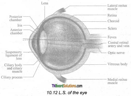
Answer:
The conjunctiva is a thin, protective mucous membrane found lining the outer surface of the eyeball.
The eye has two compartments, the anterior and posterior compartments. The anterior compartment has two chambers, the first one lies between the cornea and iris and the second one lies between the iris and lens. These two chambers are filled with a watery fluid called aqueous humor. The posterior compartment lies between the lens and retina and it is filled with a jelly-like fluid called vitreous humor that helps to retain the Spherical nature of the eye. The eye lens is transparent and biconvex, made up of long columnar epithelial cells called lens fibers.
The eyeball is spherical in nature. The wall of the eyeball consists of three layers: fibrous Sclera, vascular Choroid, and sensory Retina.
The outer coat is composed of dense non-vascular connective tissue. It has two regions: the anterior cornea and the posterior sclera. The cornea is a non-vascular transparent coat formed of stratified squamous epithelium.
Sclera forms the white of the eye and protects the eyeball. Posteriorly the sclera is innervated by the optic nerve.
The choroid is a highly vascularized pigmented layer that nourishes all the eye layers and its pigments absorb light to prevent internal reflection.
Anteriorly the choroid thickens to form the ciliary body and iris. Iris is the colored portion of the eye lying between the cornea and lens. The aperture at the center of the iris is the pupil through which the light enters the inner chamber.
Iris is made of two types of muscles the dilator papillae (the radial muscle) and the sphincter papillae (the circular muscle).In the bright light, the circular muscle in the iris contract; so that the size of the pupil decreases and less light enters the eye. In the dim light, the radial muscle in the iris contract; so that the pupil size increases and more light enters the eye. Smooth muscle present in the ciliary body is called the ciliary muscle which alters the convexity of the lens for near and far vision. The ability of the eyes to focus objects at varying distances is called accommodation which is achieved by suspensory ligament, ciliary muscle, and ciliary body. The suspensory ligament extends from the ciliary body and helps to hold the lens in its upright position. The ciliary body is provided with blood capillaries that secrete a watery fluid called aqueous humor that fills the anterior chamber.
The retina forms the innermost layer of the eye. The neural retina layer contains three types of cells: photoreceptor cells – cones and rods, bipolar cells, and ganglion cells. The yellow flat spot at the center of the posterior region of the retina is called macula lutea which is responsible for sharp detailed vision. A small depression present in the center of the yellow spot is called fovea centralis which contains only cones. The optic nerves and the retinal blood vessels enter the eye slightly below the posterior pole, which is devoid of photoreceptors; hence this region is called a blind spot.
Question 38.
Write the mechanism involved in the vision process.
Answer:
When light enters the eyes, it gets refracted by the cornea, aqueous humor, and lens and it is focused on the retina and excites the rod and cone cells. The photopigment consists of Opsin, the protein part, and Retinal, a derivative of vitamin A. Light induces dissociation of retinal from opsin and causes the structural changes in opsin. This generates an action potential in the photoreceptor cells and is transmitted by the optic nerves to the visual cortex of the brain, via bipolar cells, ganglia, and optic nerves, for the perception of vision.
Question 39.
Arun cannot able to see the nearby objects clearly. What is his problem with his eye called? How this condition occurs and how can it be rectified?
Answer:
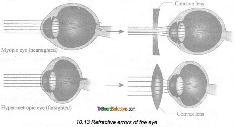
Myopia.
The affected person can see the nearby objects but not the distant objects. This condition may result due to an elongated eyeball or thickened lens; so that the image of a distant object is formed in front of the yellow spot. This error can be corrected using a concave lens that diverges the entering light rays and focuses it on the retina.
![]()
Question 40.
What is long-sightedness? What is the condition of the eye in this case? How can it be rectified?
Answer:
Hypermetropic (long-sightedness): the affected person can see only the distant objects clearly but not the objects nearby. This condition results due to a shortened – eyeball and thin lens; so the image of the closest object is converged behind the retina. This defect can be overcome by using a convex lens that converges the entering light rays on the retina.
Question 41.
Differentiate the rod and cone cells of the eyes.
Answer:
| Rod cells | Cone cells |
| Rods are responsible for vision in dim light. | The cones are responsible for color vision and work best in bright light. |
| The pigment present in the rods is rhodopsin, formed of a protein scotopsin and retinal (an aldehyde of vitamin A). | The pigment present in the cones is photopsin, formed of opsin protein and retinal. |
| There are about 120 million rod cells. | There may be 6-7 million cone cells. |
| Rods are predominant in the extra fovea region. | Cones are concentrated in the fovea region. |
Question 42.
Write few lines about the eye lens.
Answer:
The eye lens is transparent and biconvex, made up of long columnar epithelial cells called lens fibers. These cells are accumulated with the proteins called crystalline.
Question 43.
What is style?
Answer:
Infection of ciliary glands of the eye by bacteria causes a painful, pus-filled swelling called a Stye.
Question 44.
What is the reason for getting “Madras eye” in human eyes?
Answer:
Dilation and congestion of the blood vessels of the conjunctiva due to local irritation or infection are the cause of bloodshot eye (conjunctivitis – commonly called Madras eye).
Question 45.
What are the visual pigments present in the cones for color vision?
Answer:
Visual pigments for color vision are
- The red cones having the visual pigment, Erythropsin is sensitive to long-wavelength close to 560 nm.
- The green cones having the pigment, chloropsin is sensitive to the medium wavelength of 530 nm.
- The blue cones having the pigment, cyanopsin is sensitive to a short-wavelength of 420 nm.
![]()
Question 46.
Write the different types of refractive errors that occur in the eye with suitable diagrams?
Answer:
Myopia (nearsightedness): The affected person can see the nearby objects but not the distant objects. This condition may result due to an elongated eyeball or thickened lens; so that the image of a distant object is formed in front of the yellow spot. This error can be corrected using a concave lens that diverges the entering light rays and focuses them on the retina.
Hypermetropia (long-sightedness): The affected person can see only the distant objects clearly but not the objects nearby. This condition results due to a shortened eyeball and thin lens; so the image of the closest object is converged behind the retina. This defect can be overcome by using a convex lens that converges the entering light rays on the retina.
Presbyopia: Due to aging, the lens loses elasticity and the power of accommodation. Convex lenses are used to correct this defect.
Astigmatism is due to the rough (irregular) curvature of the cornea or lens. Cylindrical glasses are used to correct this error.
Cataract: Due to the changes in the nature of the protein, the lens becomes opaque. It can be corrected by surgical procedures.
Question 47.
Name the parts of the organ of equilibrium involved in the following functions.
Answer:
- Linear movement of the body – Maculae
- Changes in the body position – Perilymph and endolymph
- Rotational movement of the head – Semicircular canals
Question 48.
Write the anatomy of the ear.
Answer:
Anatomically, the ear is divided into three regions: the external ear, the middle ear, and the internal ear.
The external ear consists of the pinna, external auditory meatus, and eardrum. The pinna is the flap of elastic cartilage covered by skin. It collects the sound waves. The external auditory meatus is a curved tube that extends up to the tympanic membrane [the ear drum]. The tympanic membrane is composed of connective tissues covered with skin outside and with mucus membrane inside.
There are very fine hairs and wax-producing sebaceous glands called ceruminous glands in the external auditory meatus. The combination of hair and ear wax [cerumen] helps in preventing dust and foreign particles from entering the ear.
The middle ear is a small air-filled cavity in the temporal bone. It is separated from the external ear by the eardrum and from the internal ear by a thin bony partition; the bony partition contains two small membrane-covered openings called the oval window and the round window.
The middle ear contains three ossicles: malleus [hammer bone], incus [anvil bone], and stapes [stirrup bone] which are attached to one another. The malleus is attached to the tympanic membrane and its head articulates with the incus which is the intermediate bone lying between the malleus and stapes. The stapes is attached to the oval window in the inner ear. The ear ossicles transmit sound waves to the inner ear. A tube called Eustachian tube connects the middle ear cavity with the pharynx. This tube helps in equalizing the pressure of air on either side of the eardrum.
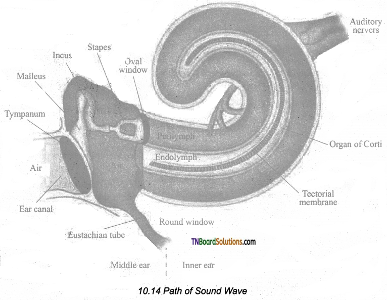
The inner ear is the fluid-filled cavity consisting of two parts, the bony labyrinth, and the membranous labyrinths. The bony labyrinth consists of three areas: cochlea, vestibule, and semicircular canals. The cochlea is a coiled portion consisting of 3 chambers namely: scala vestibuli and scala tympani- these two are filled with perilymph; and the scala media is filled with endolymph. At the base of the cochlea, the scala vestibule ends at the ‘oval window’ whereas the scala tympani ends at the ‘round window’ of the middle ear. The chambers scala vestibuli and scala media are separated by a membrane called Reisner’s membrane whereas the scala media and scala tympani are separated by a membrane called Basilar membrane.
Organ of Corti The organ of Corti is a sensory ridge located on the top of the Basilar membrane and it contains numerous hair cells that are arranged in four rows along the length of the basilar membrane. Protruding from the apical part of each hair cell is hair-like structures known as stereocilia. During the conduction of sound waves, stereocilia makes a contact with the stiff gel membrane called the tectorial membrane, a roof-like structure overhanging the organ of Corti throughout its length.
![]()
Question 49.
Give an account bn the mechanism of hearing.
Answer:
Sound waves entering the external auditory meatus fall on the tympanic membrane. This causes the eardrum to vibrate, and these vibrations are transmitted to the oval window through the three auditory ossicles. Since the tympanic membrane is 17-20 times larger than the oval window, the pressure exerted on the oval window is about 20 times more than that on the tympanic membrane. This increased pressure generates pressure waves in the fluid of perilymph. This pressure causes the round window to alternately bulge outward and inward meanwhile the basilar membrane along with the organ of Corti move up and down. These movements of the hair alternately open and close the mechanically gated ion channels in the base of hair cells and the action potential is propagated to the brain as sound sensation through the cochlear nerve.
Question 50.
Write clown the various defects of the ear.
Answer:
Deafness may be temporary or permanent. It can be further classified into conductive deafness and sensory-neural deafness. Possible causes for conductive deafness may be due to
- The blockage of the ear canal with earwax,
- Rupture of the eardrum.
- Middle ear infection with fluid accumulation.
- Restriction of ossicular movement.
In sensory-neural deafness, the defect may be in the organ of the Corti or the auditory nerve or in the ascending auditory pathways or auditory cortex.
Question 51.
Explain, the structure of the “Organ of equilibrium”.
Answer:
Balance is part of a sense called proprioception, which is the ability to sense the position, orientation, and movement of the body. The organ of balance is known as the vestibular system which is located in the inner ear next to the cochlea. The vestibular system is composed of a series of fluid-filled sacs and tubules. These sacs and tubules contain endolymph and are kept in the surrounding perilymph. These two fluids, perilymph, and endolymph, respond to the mechanical forces, during changes occurring in body position and acceleration.
The utricle and saccule are two membranous sacs, found nearest the cochlea, and contain equilibrium receptor regions called maculae that are involved in detecting the linear movement of the head. The maculae contain the hair cells that act as mechanoreceptors. These hair cells are embedded in a gelatinous otolithic membrane that contains small calcareous particles called otoliths.
The canals that lie posterior and lateral to the vestibule are semicircular canals; they are anterior, posterior, and lateral canals oriented at right angles to each other. At one end of each semicircular canal, at its lower end has a swollen area called the ampulla. Each ampulla has a sensory area known as crista ampullar which is formed of sensory hair cells and supporting cells. The function of these canals is to detect the rotational movement of the head.
![]()
Question 52.
Write down the receptors present in the skin.
Answer:
Some of the sensory receptors present in the skin are –
- Tactile Merkel disc is a light touch receptor lying in the deeper layer of the epidermis.
- Hair follicle receptors are light-touch receptors lying around the hair follicles.
- Meissner’s corpuscles are small light pressure receptors found just beneath the epidermis in the dermal papillae. They are numerous in hairless skin areas such as fingertips and soles of the feet.
- Pacinian corpuscles are the large egg-shaped receptors found scattered deep in the dermis and monitoring vibration due to pressure. It allows detecting different textures, temperatures, hardness, and pain.
- Ruffini endings that lie in the dermis respond to continuous pressure.
- Krause end bulbs are thermoreceptors that sense temperature.
Question 53.
What is vitiligo? What are its symptoms?
Answer:
Vitiligo (Leucoderma) is a condition in which the melanin pigment is lost from areas of the skin, causing white patches, often with no clear cause. Vitiligo is not contagious. It can affect people of any age, gender, or ethnic group. The patches appear when melanocytes fail to synthesis melanin pigment.
Question 54.
What is spike potential?
Answer:
Due to the rapid influx of Na+ ions, the membrane potential shoots rapidly up to +45mV in the neuron, which is called the spike potential.
Question 55.
Define threshold potential and threshold stimulus.
Answer:
During depolarization, when enough Na+ ions enter the cell, the action potential reaches a certain level, called threshold potential [-55mV]. The particular stimulus which is able to bring the membrane potential to the threshold is called threshold stimulus.
![]()
Question 56.
Define the following.
Answer:
(i) Leakage channels.
(ii) Ligand-gated channels.
(iii) Voltage-gated channels.
- Leakage Channels are ionic channels that remain open all the time.
- Ligand-gated channels are chemically gated channels that open or close in response to chemical stimuli.
- Voltage-gated channels are mechanically gated channels that open in response to a physical stimulus in the form of vibration such as touch and pressure.
Question 57.
What are actions take place during Ligand-gated channels?
Answer:
Ligand-gated channels are located between the presynaptic membrane of the first axon and postsynaptic membrane of the cell body of the second neuron [i.e. dendrites and cell bodies]. The neurotransmitter acetylcholine opens ligand channels that allow Na+ and Ca++ ions to diffuse inward and K+ ions diffuse outward.
Question 58.
How potential difference across the axolemma is maintained during leakage channels.
Answer:
K+ leakage channels are more in number than the Na+ leakage channels. Sarcolemma has greater permeability to K+ ions than Na+ ions. These ions keep moving continuously maintain the potential difference across the axolemma.
Question 59.
Name the two types of voltage-gated channels.
Answer:
There are two types of voltage-gated channels.
- Sodium voltage-gated channel
- Potassium voltage-gated channel.
![]()
Choose the correct answer.
1. The structural and functional units of the neural system are:
(a) nephrons
(b) neurons
(c) neuroglia
(d) intemeurons
Answer:
(b) neurons
2. The non-nervous special cells which forms the supporting cells of the nervous tissue are called:
(a) afferent neurons
(b) efferent neurons
(c) neuroglia
(d) dendrons
Answer:
(c) neuroglia
3. The plasma membrane covering the neuron is called:
(a) cell wall
(b) primary wall
(c) neurilemma
(d) axolemma
Answer:
(c) neurilemma
4. The area from where the axon arises from the cell body of the neuron is called:
(a) Schwann cells
(b) Node of ranvier
(c) Nissil body
(d) Axon hillock
Answer:
(d) Axon hillock
![]()
5. Nissil’s granules are absent in the ……….. area of the neuron.
(a) dendrites
(b) cell body
(c) axon
(d) myelin sheath
Answer:
(c) axon
6. The longest cells in the human body are the:
(a) nephrons
(b) axons
(c) dendrons
(d) neurons
Answer:
(d) neurons
7. ………… is the longest axon in the human body.
(a) Vagus nerve
(b) Cervical nerve
(c) Sciatic nerve
(d) Sacral nerve
Answer:
(c) Sciatic nerve
8. The axon of the peripheral nerves is surrounded by:
(a) nodes of ranvier
(b) nissil’s bodies
(c) axon hillock
(d) schwann cells
Answer:
(d) schwann cells
![]()
9. The gaps in the myelin sheath of the axon in between the adjacent Schwann cells are called:
(a) synaptic vesicle
(b) synaptic knob
(c) nodes of ranvier
(d) neuromuscular junction
Answer:
(c) nodes of ranvier
10. In neurons, the normal value of resting membrane potential is:
(a) -70mV
(b) -40m V
(c) -50mV
(d) -17mV
Answer:
(a) -70mV
11. When a nerve fiber is in the stimulated stage the following action will result:
(a) Sodium voltage-gated opens
(b) Potassium voltage-gated opens
(c) Sodium voltage-gated closes
(d) Ligand-gated opens
Answer:
(a) Sodium voltage-gated opens
12. If repolarization becomes more negative than the resting potential -70mV to about -90mV, it is called:
(a) depolarization
(b) repolarization
(c) hyperpolarization
(d) hyperpolarization
Answer:
(c) hyperpolarization
![]()
13. Nerve impulses travel at the speed of:
(a) 1-3 m/s
(b) 1-300 m/s
(c) 2-250 m/s
(d) 1-310 m/s
Answer:
(b) 1-300 m/s
14. The junction between two neurons is called a:
(a) synapse
(b) nodes of Ranvier
(c) synaptic cleft
(d) septum pellucidum
Answer:
(a) synapse
15. A small gap between the pre and postsynaptic membrane is called:
(a) synaptic cleft
(b) synaptic vesicle
(c) synaptic knob
(d) synapse
Answer:
(a) synaptic cleft
16. The synaptic vesicles of the axon terminal is filled with:
(a) neurotransmitters
(b) cerebrospinal fluid
(c) plasma
(d) mucus
Answer:
(a) neurotransmitters
17. The outer dura mater and the median Arachnoid membranes of the. the brain has separated from each other by means of space called:
(a) dural space
(b) arachnoid space
(c) subarachnoid space
(d) subdural space
Answer:
(d) subdural space
![]()
18. ……….. part of the brain is called “Seat of intelligence” and forms the major part of the brain.
(a) Cerebrum
(b) Cerebellum
(c) Diencephalon
(d) Hypothalamus
Answer:
(a) Cerebrum
19. The cerebral hemispheres of the cerebrum are connected by a tract of nerve fibers called:
(a) infundibulum
(b) corpus callosum
(c) cauda equina
(d) choroid plexus
Answer:
(b) corpus callosum
20. ……….. serve as a relay center for impulses between the spinal cord, brainstem, and cerebrum.
(a) Hypothalamus
(b) Pineal body
(c) Thalamus
(d) Infundibulum
Answer:
(c) Thalamus
21. ……….. is the major coordinating center for sensory and motor signaling.
(a) Olfactory bulbs
(b) Brainstem
(c) Thalamus
(d) Corpora quadrigemina
Answer:
(c) Thalamus
![]()
22. The hypothalamus contains a pair of the small rounded bodies called ……….. which are involved in olfactory reflexes and emotional responses to odor.
(a) corpora quadrigemina
(b) mamillary bodies
(c) foramen of Monro
(d) hippocampus
Answer:
(b) mamillary bodies
23. ………. system of our body is called the “emotional brain.”
(a) Neural system
(b) Muscular system
(c) Sensory receptor system
(d) Limbic system
Answer:
(d) Limbic system
24. The four rounded bodies in the dorsal portion of the midbrain is called:
(a) corpora quadrigemina
(b) mamillary bodies
(c) olfactory bulbs
(d) cauda equina
Answer:
(a) corpora quadrigemina
25. ……….. is the reflex center of the brain for vision and hearing.
(a) Cerebral peduncles
(b) Septum pellucidum
(c) Corpora quadrigemina
(d) Choroid plexus
Answer:
(c) Corpora quadrigemina
![]()
26. ………… is the second largest part of the brain.
(a) Cerebrum
(b) Medulla oblongata
(c) Cerebellum
(d) Pons varoli
Answer:
(c) Cerebellum
27. ………… part of the brain controls and coordinates the muscular movements and body equilibrium.
(a) Cerebrum
(b) Hypothalamus
(c) Vermis
(d) Cerebellum
Answer:
(d) Cerebellum
28. Cardiovascular reflexes, respiration, and gastric secretions are controlled by:
(a) Medulla oblongata
(b) Interventricular foramen.
(c) Cerebral hemispheres
(d) Cerebellum
Answer:
(a) Medulla oblongata
29. The 1st and the 2nd ventricles of the brain are communicated with the 3rd ventricle through an opening called:
(a) aqueduct of Sylvius
(b) foramen of Monro
(c) intervertebral foramina
(d) pupil
Answer:
(b) foramen of Monro
![]()
30. The thick bundle of elongated nerve roots within the lower vertebral canal is called the:
(a) choroid plexus
(b) cauda equina
(c) peripheral neural, system
(d) intervertebral foramina
Answer:
(b) cauda equina
31. There are ……….. pairs of cranial nerves that arise from the brain.
(a) 11
(b) 12
(c) 13
(d) 14
Answer:
(b) 12
32. There are ……….. pairs of spinal nerves emerge out from the spinal cord.
(a) 30
(b) 29
(c) 31
(d) 13
Answer:
(c) 31
33. The spinal nerves emerge out from the spinal cord through spaces called:
(a) foramen of Monro
(b) intervertebral foramina
(c) aqueduct of sylvium
(d) synapse
Answer:
(b) intervertebral foramina
![]()
34. …………. located in the upper lateral region of each orbit of secrete tears.
(a) Sebaceous glands
(b) Lacrymal glands
(c) Meibomian glands
(d) Oil glands
Answer:
(b) Lacrymal glands
35. The excess of aqueous humor in an eye drains out through:
(a) foramen of Monro
(b) canal of schlemm
(c) fovea centralis
(d) macula lutea
Answer:
(b) canal of schlemm
36. ……….. layer of an eye is the highly vascularized pigmented layer.
(a) Sclera
(b) Choroid
(c) Retina
(d) Eyelids
Answer:
(b) Choroid
37. The normal value of intraocular pressure of an eye is:
(a) 16 mmHg
(b) 61 mmHg
(c) 22 mmHg
(d) 6 mmHg
Answer:
(a) 16 mmHg
38. The convexity of the lens of an eye for near and far vision is altered by:
(a) sphincter muscle
(b) ciliary muscle
(c) suspensory ligaments
(d) dilator papillae
Answer:
(b) ciliary muscle
![]()
39. ………. is responsible for sharp detailed vision of eye, which is present in the centre of the posterior region of retina.
(a) Macula lutea
(b) Fovea centralis
(c) Pupil
(d) Canal of Schlemm
Answer:
(a) Macula lutea
40. The pigmenf present in the cone cells of an eye is:
(a) anthocyanin
(b) carotenoids
(c) phytochrome
(d) photopsin
Answer:
(d) photopsin
41. Due to aging, the lens of an eye looses its elasticity and the power of accommodation, what is the condition called?
(a) Astigmatism
(b) Myopia
(c) Presbyopia
(d) Hypermetropia
Answer:
(c) Presbyopia
42. There are about ………. million-rod cells in the human eye.
(a) 1200
(b) 120
(c) 12
(d) 102
Answer:
(b) 120
![]()
43. Match:
| 1. Myopia | (a) Lens loses elasticity |
| 2. Hypermetropia | (b) The lens becomes opaque |
| 3. Presbyopia | (b) Rough curvature of cornea or lens |
| 4. Astigmatism | (c) Short sightedness |
| 5. Cataract | (d) Long sightedness |
(a) 1 -(d), 2-(e), 3-(a), 4-(c), 5-(b)
(b) 1-(e), 2-(d),3-(c), 4-(a), 5-(b)
(c) 1-(b), 2-(a),3-(c), 4-(d),5-(e)
(d) 1-(d), 2-(a), 3-(e), 4-(c), 5-(b)
Answer:
(a) 1 -(d), 2-(e), 3-(a), 4-(c), 5-(b)
44. The wax glands present in an ear is called:
(a) sebaceous glands
(b) meibomian glands
(c) oil glands
(d) ceruminous glands
Answer:
(d) ceruminous glands
45. The hair cells of the basilar membrane of the inner ear has hair-like projections called:
(a) cilia
(b) flagella
(c) stereocilia
(d) ciliary epithelium
Answer:
(c) stereocilia
46. The intensity of sound is measured in:
(a) mV
(b) decibels
(c) kelvin
(d) mole
Answer:
(b) decibels
![]()
47. The receptors for taste and smell are called:
(a) chemoreceptors
(b) mechanoreceptors
(c) tactile receptors
(d) phono receptors
Answer:
(a) chemoreceptors
48. The tongue has many small projections called ……….. which give the tongue an abrasive feel.
(a) papillae
(b) dilator papillae
(c) sphincter papillae
(d) ruffini endings
Answer:
(a) papillae
49. ………. are the small light pressure receptors found numerous in hairless skin areas such as fingertips and soles of the feet.
(a) Pacinian corpuscles
(b) Meissner’s corpuscles
(c) Krause end bulbs
(d) Tactile Merkel disc
Answer:
(b) Meissner’s corpuscles
50. ………… are the thermoreceptors found on the skin that sense temperature.
(a) Gustatory epithelial cells
(b) Olfactory receptor cells
(c) Krause end bulbs
(d) Pacinian corpuscles
Answer:
(c) Krause end bulbs
![]()
51. Match the following:
| 1. Nerve cell | (a) Neurilemma, axolemma |
| 2. Plasma membrane | (b) Nissl’s granules |
| 3. Cytoplasm | (c) Neuron |
| 4. Endoplasmic reticulum | (d) Retina of the eye |
| 5. Bipolar neurons | (e) Neuroplasm |
(a) 1-(d), 2-(a), 3-(c), 4-(e), 5-(b)
(b) 1-(a), 2-(c), 3-(d), 4-(b), 5-(e)
(c) 1-(d), 2-(e), 3-(c), 4-(a), 5-(b)
(d) 1-(c), 2-(a), 3-(e), 4-(b), 5-(d)
Answer:
(d) 1-(c), 2-(a), 3-(e), 4-(b), 5-(d)